face Archives - Page 3 of 6 - Dr. Walter Zamarian Jr.

Categoria: face

  • Non-Surgical vs Surgical Rhinoplasty: Limits, Risks and Choices

    Non-Surgical vs Surgical Rhinoplasty: Limits, Risks and Choices

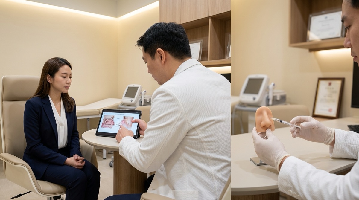
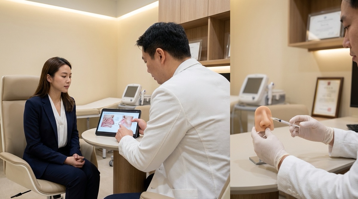
    Non-surgical rhinoplasty can camouflage selected minor nasal contour issues by adding filler, while surgical rhinoplasty is required when the goal is reduction, cartilage or bone reshaping, septal correction, valve support or breathing improvement. The safer choice depends on anatomy, goals, medical history, prior filler, airway function and tolerance for recovery.

    Many patients ask whether a nose can be improved without surgery. The honest answer is sometimes yes, but only within narrow limits. A filler can add volume to disguise a small irregularity; it cannot make a large nose smaller, narrow a wide tip, remove bone, rebuild cartilage or correct a deviated septum.

    Medical review

    Written and reviewed by Dr. Walter Zamarian Jr., plastic surgeon in Londrina, Brazil. CRM-PR 17.388, RQE 15.688, full member of the Brazilian Society of Plastic Surgery (SBCP) and member of the American Society of Plastic Surgeons (ASPS). 20+ years of experience and 8,000+ surgeries performed. Last reviewed: May 24, 2026.

    What non-surgical rhinoplasty can do

    Non-surgical rhinoplasty, also called a liquid rhinoplasty or nasal filler treatment, usually uses hyaluronic acid filler to change the way light reflects on the nose. It can soften a small depression, camouflage a mild dorsal irregularity, improve selected asymmetries or create the impression of a smoother profile.

    The key limitation is that filler adds volume. It does not remove a hump, reduce size, refine cartilage, narrow thick skin, support weak nasal valves or improve breathing. If the nose already feels too large, adding filler may make the face look less balanced even if the profile line appears smoother.

    For a deeper discussion of filler limits and complications, see the related guide on non-surgical rhinoplasty limits and filler risks.

    What surgical rhinoplasty can do

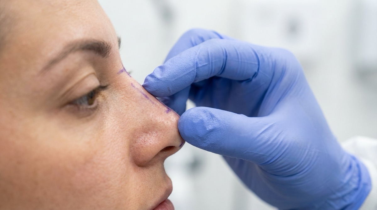
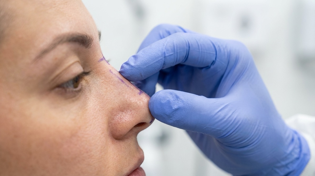
    Surgical rhinoplasty changes nasal structure. Depending on the case, it can refine the tip, adjust the bridge, reduce or support the dorsum, straighten selected deformities, use cartilage grafts, improve nasal valve support and address breathing problems when combined with septal or functional work.

    In my practice, I think structurally: the nose must look balanced and remain supported over time. In selected patients, planning may include ultrasonic rhinoplasty, septorhinoplasty or, when prior surgery has already altered the anatomy, revision rhinoplasty.

    Surgery can be long-lasting, but it should not be described as a fixed or guaranteed result. Healing, scar behavior, skin thickness, cartilage memory, trauma, aging and individual anatomy all influence the final outcome.

    Practical comparison

    FactorNon-surgical rhinoplastySurgical rhinoplasty
    Main mechanismAdds filler to camouflage selected contour issuesChanges cartilage, bone, septum, support and shape
    Can reduce nose size?NoYes, in selected anatomical patterns
    Can improve breathing?NoSometimes, when functional anatomy is addressed
    Best useSmall camouflage, mild asymmetry, selected refinementStructural change, reduction, tip work, septum/valve support
    RecoveryUsually shorter, but swelling/bruising can occurLonger recovery with swelling that evolves over months
    ReversibilityMany hyaluronic acid fillers can be treated with hyaluronidaseCorrection requires healing time and, sometimes, revision surgery
    Main risksVascular occlusion, necrosis, visual symptoms, nodules, migrationAnesthesia, bleeding, infection, breathing change, asymmetry, revision

    When filler may be reasonable

    Nasal filler may be reasonable when the concern is small, cosmetic and caused by a contour depression that can be safely camouflaged. It may also help selected patients who want a temporary change, or selected post-rhinoplasty patients who need a minor contour refinement and are not candidates for revision surgery at that time.

    The indication must be conservative. The nose is a high-risk injection area because its blood supply connects with vessels around the eye. The person injecting must understand nasal vascular anatomy, have an emergency plan, and have hyaluronidase immediately available when hyaluronic acid filler is used.

    When surgery is usually the more honest discussion

    Surgery becomes the more honest discussion when the patient wants a smaller nose, a more defined tip, correction of a significant hump, better symmetry after trauma, correction of a deviated septum, improvement in nasal valve support or a plan that addresses both appearance and breathing.

    Repeated filler can delay the right decision and may distort tissue planes. If filler has been placed many times, I evaluate whether it should be dissolved before surgery, whether it has caused fibrosis or irregularity, and whether the surgical plan needs to be adjusted.

    Filler risks patients should understand

    Dermal filler is a medical procedure. The most serious complication is vascular occlusion, when filler enters or compresses a blood vessel. This can reduce blood flow and cause severe pain, skin blanching, mottled color change, blisters, skin necrosis, scarring and, rarely, visual symptoms, blindness or stroke.

    Other possible filler complications include bruising, swelling, infection, nodules, Tyndall effect, migration, delayed inflammatory reaction, asymmetry and dissatisfaction with shape. The fact that hyaluronic acid can often be dissolved does not make the procedure casual or without risk.

    Surgical rhinoplasty risks patients should understand

    Rhinoplasty is surgery. Possible risks include anesthesia-related problems, bleeding, infection, poor wound healing, scarring, prolonged swelling, numbness, skin suffering or necrosis, septal perforation, breathing changes, asymmetry, contour irregularity, dissatisfaction with appearance and the possibility of revision surgery.

    Risk reduction starts before the operating room: correct indication, medical history review, smoking and nicotine avoidance, realistic expectations, careful photography, functional nasal assessment and a clear discussion of what surgery can and cannot change.

    Warning signs after filler or surgery

    After nasal filler, urgent warning signs include severe or increasing pain, white or mottled skin, new blisters, darkening skin, eye pain, blurred vision, loss of vision or neurological symptoms. After rhinoplasty, warning signs include fever, pus, rapidly worsening swelling, heavy bleeding, chest pain, shortness of breath, calf swelling or severe uncontrolled pain. These symptoms require immediate contact with the treating physician or emergency care.

    Frequently asked questions

    Can nose filler replace rhinoplasty?

    Nose filler cannot replace rhinoplasty when the goal is reduction, tip refinement, cartilage reshaping, septal correction or breathing improvement. It can only camouflage selected minor contour issues by adding volume.

    Is non-surgical rhinoplasty safer because it avoids surgery?

    Non-surgical rhinoplasty avoids surgical recovery, but it is not automatically safer. Nasal filler carries rare but serious vascular risks, including skin necrosis, visual symptoms, blindness or stroke.

    Can filler make future rhinoplasty harder?

    Filler can make future rhinoplasty more complex when it leaves residual product, inflammation, fibrosis or distorted planes. A surgeon may recommend waiting, dissolving hyaluronic acid filler or adjusting the surgical plan.

    How do I choose between filler and surgery?

    The choice depends on what bothers you anatomically. If the issue is minor camouflage, filler may fit; if the issue is size, cartilage, bone, septum, nasal valves or breathing, surgical rhinoplasty is usually the more appropriate conversation.

    How I approach the decision in consultation

    During consultation in Londrina, I examine the nose from the front, profile, base and functional perspective. I look at skin thickness, tip support, dorsum, septum, nasal valves, prior filler, prior surgery, breathing symptoms and facial proportions. The aim is not to push a procedure, but to match the treatment to the anatomy.

    For related reading, see the pages on rhinoplasty, ultrasonic rhinoplasty, septorhinoplasty, revision rhinoplasty and facial fillers, as well as the safety guide for structural rhinoplasty in Brazil for international patients.

  • Revision Rhinoplasty: When a Second Nose Surgery Makes Sense

    Revision Rhinoplasty: When a Second Nose Surgery Makes Sense

    Revision rhinoplasty is considered after the nose has healed when persistent breathing problems, structural collapse, significant asymmetry or a clearly correctable contour problem remains; many patients should wait 12 to 18 months before deciding. A second nose surgery can help selected patients, but not every dissatisfaction should be treated with another operation.

    Patients often seek revision because they still cannot breathe well, the nose looks over-operated, the tip lost support, the bridge became irregular, or the result does not match what they expected. The first step is not to schedule surgery. The first step is to understand whether there is an objective, correctable problem and whether the tissues are ready for another operation.

    Medical review

    Written and reviewed by Dr. Walter Zamarian Jr., plastic surgeon in Londrina, Brazil. CRM-PR 17.388, RQE 15.688, full member of the Brazilian Society of Plastic Surgery (SBCP) and member of the American Society of Plastic Surgeons (ASPS). 20+ years of experience and 8,000+ surgeries performed. Last reviewed: May 24, 2026.

    What is revision rhinoplasty?

    Revision rhinoplasty, also called secondary rhinoplasty, is nasal surgery performed after a previous rhinoplasty. It may be aesthetic, functional or both. Some cases involve a small contour issue; others require reconstruction of support that was weakened, removed or distorted during the first procedure.

    The operation is different from a primary rhinoplasty because the surgeon is working in tissue that has already healed with scars. Normal planes may be altered, cartilage may be missing, the septum may already have been used, and skin may be less forgiving. This is why planning is more important than speed.

    When a second surgery may be appropriate

    Revision may be appropriate when a patient has a problem that is visible, functional, stable and likely to improve with surgery. Examples include persistent nasal obstruction, nasal valve collapse, residual or recurrent septal deviation, a crooked nose, an irregular dorsum, tip collapse, alar retraction, nostril asymmetry, inverted-V deformity or structural weakness that continues to worsen over time.

    Aesthetic dissatisfaction alone is not enough. I need to determine whether the concern is surgically correctable, whether the expected improvement is meaningful, and whether the risk of another operation is justified. Sometimes the safest recommendation is to wait, treat the airway medically, dissolve residual filler, or not operate.

    When it is too early to revise

    In most patients, I prefer waiting 12 to 18 months after the previous rhinoplasty before deciding on revision. Swelling can last a long time, especially in the nasal tip, thick skin, revision cases and patients who had extensive structural work. A concern that looks important at month four may become much less relevant by month twelve.

    Earlier evaluation is important when there is infection, severe obstruction, trauma, progressive collapse, exposed graft, skin suffering or a rapidly worsening deformity. Evaluation does not always mean immediate surgery; it means the problem should be assessed promptly and followed closely.

    Why revision rhinoplasty is more complex

    Scar tissue changes the anatomy

    Every rhinoplasty creates scar tissue. In revision, the tissue planes are less distinct, bleeding can be less predictable and the skin envelope may not redrape as easily. This can limit how much refinement is possible.

    Cartilage may be missing or weakened

    Primary rhinoplasty sometimes removes or weakens cartilage. If support is insufficient, revision is not only about contour; it may require rebuilding the framework of the nose. This is especially important when breathing is affected or when the tip, middle vault or nostril rims have lost support.

    Expectations must be narrower

    A revision plan must respect what the previous surgery left behind. The goal may be improvement in breathing, better support, a smoother bridge or a more balanced shape, not a perfect or completely new nose. Clear limits protect the patient from unnecessary surgery.

    Cartilage graft options

    Revision rhinoplasty often requires grafting. The best graft source depends on how much cartilage is needed, what remains from the septum, whether the ear is suitable and whether a stronger graft is required.

    • Septal cartilage: useful when enough remains, but it may have been removed or altered during the first operation.
    • Ear cartilage: useful for selected contour and rim support, but it is curved and limited in quantity.
    • Rib cartilage: useful for major reconstruction, severe support loss or multiple previous surgeries, but it adds donor-site pain, scar, possible warping, possible visibility/palpability and rare chest-wall complications.

    Rib cartilage can be a valuable tool, but it should not be presented as simple or automatically necessary. The indication must be individualized. When the airway and structure need major support, rib cartilage may be the safest source of enough material; in smaller revisions, septal or ear cartilage may be enough.

    Functional revision: breathing matters

    A revision consultation must evaluate breathing, not only appearance. Persistent obstruction can be related to residual septal deviation, nasal valve collapse, turbinate issues, synechiae, scarring or loss of structural support. In selected patients, revision may overlap with septorhinoplasty because form and function are connected.

    When bone work is needed, ultrasonic rhinoplasty can be considered in selected cases, but technology is not the plan. The plan is anatomical: identify the cause, rebuild support when needed and avoid weakening the nose further.

    Recovery after revision rhinoplasty

    Recovery after revision can resemble primary rhinoplasty, but swelling often lasts longer. The tip may take many months to refine, and scarred tissues can feel firmer during healing. If rib cartilage is harvested, the chest donor site also needs recovery time and pain control.

    Patients should expect staged healing rather than an early final answer. The first weeks are about splint care, bruising and swelling. The following months are about gradual definition. The final judgment may require 12 to 18 months, especially in thick skin or complex revisions.

    Risks and red flags

    Revision rhinoplasty carries the risks of rhinoplasty plus the added complexity of previous surgery. Possible risks include anesthesia-related problems, bleeding, infection, poor healing, scarring, prolonged swelling, numbness, skin suffering or necrosis, septal perforation, breathing changes, asymmetry, contour irregularity, graft warping, graft visibility or palpability, donor-site pain or scar, dissatisfaction and the possibility of further surgery.

    Urgent red flags include fever, pus, rapidly worsening swelling, heavy bleeding, severe uncontrolled pain, worsening obstruction, skin color change, chest pain, shortness of breath or calf swelling. These signs require immediate contact with the treating surgeon or emergency care.

    Expectations and emotional readiness

    Revision rhinoplasty is emotionally charged. Patients may feel disappointed, anxious or distrustful after the first operation. Those feelings are understandable, but they must be separated from surgical decision-making. If distress is disproportionate to objective findings, if the concern keeps changing, or if the goal is a perfectly symmetrical nose, surgery may not be the right next step.

    My role is to explain what can realistically improve, what probably cannot, and what could become worse. A responsible consultation sometimes ends with a plan to wait, observe and photograph rather than operate immediately.

    Frequently asked questions

    How long should I wait before revision rhinoplasty?

    Most patients should wait 12 to 18 months before deciding on revision rhinoplasty, because swelling and scar maturation can change the apparent result. Earlier evaluation is appropriate for severe obstruction, infection, trauma, skin problems or progressive collapse.

    Is revision rhinoplasty riskier than primary rhinoplasty?

    Revision rhinoplasty is usually more complex because scar tissue, altered anatomy and missing cartilage reduce predictability. It can still be appropriate, but the risk-benefit discussion must be stricter than in a first rhinoplasty.

    Will I need rib cartilage?

    Rib cartilage is not needed in every revision rhinoplasty. It is considered when septal or ear cartilage is insufficient and the nose needs stronger structural support, especially after major collapse or multiple previous surgeries.

    Can revision rhinoplasty improve breathing?

    Revision rhinoplasty can improve breathing in selected patients when obstruction is caused by correctable structural problems such as septal deviation, nasal valve collapse, synechiae or loss of support. It cannot guarantee normal breathing in every case.

    How I evaluate revision candidates

    During consultation, I review the patient’s timeline, photographs, prior operative information when available, breathing symptoms, skin thickness, cartilage support, nostril shape, dorsum, tip, septum and expectations. For international patients, an online consultation may help with preliminary planning, but an in-person examination in Londrina is necessary before surgery.

    For related reading, see the pages on revision rhinoplasty, structural rhinoplasty, septorhinoplasty, ultrasonic rhinoplasty and ethnic rhinoplasty, plus the blog guides on surgical vs non-surgical rhinoplasty and structural rhinoplasty in Brazil for international patients.

  • Rinoplastia de Revisão: quando uma segunda cirurgia faz sentido

    Rinoplastia de Revisão: quando uma segunda cirurgia faz sentido

    A rinoplastia de revisão pode ser considerada depois que o nariz cicatrizou adequadamente, geralmente após 12 a 18 meses, quando permanecem dificuldade respiratória, colapso estrutural, assimetria relevante ou uma alteração de contorno claramente corrigível. Uma segunda cirurgia nasal pode ajudar pacientes selecionados, mas nem toda insatisfação depois de uma rinoplastia deve ser tratada com nova operação.

    O primeiro passo não é decidir pela cirurgia. O primeiro passo é entender se existe um problema objetivo, se ele é corrigível, se os tecidos já amadureceram e se o risco de operar novamente é justificado. Em alguns casos, a melhor conduta é observar por mais tempo; em outros, investigar a respiração, revisar exames ou planejar reconstrução estrutural.

    Revisão médica

    Texto escrito e revisado pelo Dr. Walter Zamarian Jr., cirurgião plástico em Londrina. CRM-PR 17.388, RQE 15.688, membro titular da Sociedade Brasileira de Cirurgia Plástica (SBCP) e membro da American Society of Plastic Surgeons (ASPS). Mais de 20 anos de experiência e mais de 8.000 cirurgias realizadas. Última revisão: 24 de maio de 2026.

    O que é rinoplastia de revisão?

    A rinoplastia de revisão, também chamada de rinoplastia secundária, é uma cirurgia nasal realizada depois de uma rinoplastia prévia. Ela pode ter objetivo funcional, estético ou ambos. Em alguns pacientes, a revisão corrige uma irregularidade pequena; em outros, exige reconstrução de suporte nasal que foi enfraquecido, removido ou distorcido na primeira cirurgia.

    Ela é diferente de uma primeira rinoplastia porque o cirurgião encontra tecidos já cicatrizados. Os planos anatômicos podem estar alterados, a cartilagem septal pode já ter sido usada, a pele pode estar menos complacente e a resposta ao inchaço pode ser mais lenta. Por isso, a revisão exige mais diagnóstico, mais planejamento e uma conversa mais rigorosa sobre limites.

    Quando uma segunda cirurgia pode fazer sentido

    A revisão pode ser apropriada quando existe um problema visível, funcional, estável e com boa chance de melhora cirúrgica. Exemplos incluem obstrução nasal persistente, colapso da válvula nasal, desvio septal residual ou recorrente, nariz torto, dorso irregular, perda de suporte da ponta, retração alar, assimetria das narinas, deformidade em V invertido ou enfraquecimento estrutural progressivo.

    A insatisfação estética isolada não é suficiente. É preciso diferenciar um problema real de cicatrização ainda em evolução, expectativa desalinhada, alteração pequena demais para justificar o risco ou quadro emocional que pode piorar com novas cirurgias. Operar um nariz que não precisa de revisão pode causar mais dano do que benefício.

    Quando ainda é cedo para revisar

    Na maioria dos pacientes, prefiro aguardar 12 a 18 meses após a rinoplastia anterior antes de indicar uma revisão. O inchaço pode durar muito tempo, principalmente na ponta nasal, em peles espessas, em casos de cirurgia extensa e em pacientes que já foram operados. Uma irregularidade que incomoda no quarto mês pode se tornar menos relevante depois de um ano.

    Existem exceções. Avaliação precoce é importante quando há infecção, obstrução respiratória intensa, trauma, colapso progressivo, exposição de enxerto, sofrimento de pele ou deformidade que piora rapidamente. Avaliar cedo não significa operar cedo; significa acompanhar o problema com segurança e decidir no momento certo.

    Por que a rinoplastia de revisão é mais complexa?

    Cicatriz interna muda os planos anatômicos

    Toda rinoplastia gera cicatriz. Na revisão, a fibrose pode tornar os tecidos mais rígidos, menos previsíveis e com planos de dissecção menos nítidos. Isso pode limitar a quantidade de refinamento possível e aumentar a importância de preservar a vascularização da pele.

    Cartilagem pode estar ausente ou enfraquecida

    Algumas rinoplastias primárias removem ou enfraquecem cartilagem. Quando o suporte é insuficiente, a revisão não é apenas uma cirurgia de contorno; ela pode exigir reconstrução da estrutura nasal. Isso é especialmente importante quando há dificuldade para respirar ou perda de sustentação da ponta, do dorso médio ou das bordas das narinas.

    As expectativas precisam ser mais estreitas

    A cirurgia anterior deixa limites reais. Em uma revisão, o objetivo pode ser melhorar a respiração, reconstruir suporte, suavizar uma irregularidade ou devolver proporção ao nariz, mas não criar um nariz perfeito ou completamente novo. Expectativa realista é parte da indicação médica.

    Como avalio função respiratória e estética

    A consulta precisa avaliar aparência e respiração. A obstrução nasal pode estar relacionada a desvio de septo residual, colapso da válvula nasal, alterações dos cornetos, sinéquias, cicatrizes internas ou perda de suporte estrutural. Em alguns casos, a revisão se aproxima de uma rinosseptoplastia, porque forma e função estão conectadas.

    Quando o problema envolve osso nasal, a rinoplastia ultrassônica pode ser considerada em pacientes selecionados. Ainda assim, tecnologia não substitui diagnóstico. A pergunta principal é anatômica: qual estrutura falhou, o que precisa ser preservado e o que precisa ser reconstruído?

    Enxertos na rinoplastia de revisão

    A revisão frequentemente exige enxertos de cartilagem. A escolha depende do que sobrou do septo, da quantidade de suporte necessária, do formato desejado e da presença de cirurgias anteriores.

    • Cartilagem septal: é útil quando ainda existe material suficiente, mas muitas vezes já foi usada na primeira cirurgia.
    • Cartilagem de orelha: pode ajudar em refinamentos e suporte de borda alar, mas tem quantidade limitada e curvatura própria.
    • Cartilagem de costela: pode ser indicada em reconstruções maiores, colapso importante ou múltiplas cirurgias prévias, mas acrescenta dor e cicatriz em área doadora, além de risco de empenamento, irregularidade, visibilidade, palpabilidade ou necessidade de nova correção.

    O enxerto de costela é uma ferramenta valiosa, mas não deve ser banalizado. Ele pode oferecer material abundante e firme para reconstrução, especialmente quando falta suporte nasal. Ao mesmo tempo, exige indicação precisa, técnica cuidadosa e explicação clara dos riscos.

    Recuperação depois de uma rinoplastia de revisão

    A recuperação pode lembrar a primeira rinoplastia, mas o inchaço costuma ser mais prolongado. A ponta nasal é a região que mais demora para definir, e tecidos já operados podem ficar endurecidos durante a cicatrização. Se houver retirada de cartilagem costal, a região torácica também precisa de cuidado e controle de dor.

    Nas primeiras semanas, o foco é cuidar da tala, controlar edema e evitar trauma. Nos meses seguintes, a definição aparece gradualmente. Em revisões, o julgamento final pode exigir 12 a 18 meses, principalmente em peles espessas ou casos reconstrutivos.

    Riscos e sinais de alerta

    A rinoplastia de revisão tem os riscos de uma rinoplastia somados à complexidade de operar tecidos já modificados. Podem ocorrer problemas anestésicos, sangramento, infecção, má cicatrização, cicatrizes, edema prolongado, dormência, sofrimento de pele, perfuração septal, alterações respiratórias, assimetrias, irregularidades de contorno, empenamento de enxerto, enxerto visível ou palpável, dor ou cicatriz em área doadora, insatisfação e necessidade de nova cirurgia.

    Febre, secreção purulenta, inchaço que piora rapidamente, sangramento intenso, dor fora do esperado, piora respiratória, mudança de cor da pele, dor no peito, falta de ar ou inchaço em panturrilha exigem contato imediato com a equipe cirúrgica ou atendimento de urgência.

    Expectativa e preparo emocional

    A revisão nasal tem uma carga emocional importante. Muitos pacientes chegam frustrados, inseguros ou com medo de repetir uma experiência ruim. Isso precisa ser acolhido, mas também separado da decisão cirúrgica. Se o incômodo muda a cada consulta, se a expectativa é simetria perfeita ou se o sofrimento é desproporcional ao achado físico, a cirurgia pode não ser o melhor próximo passo.

    Uma indicação responsável pode terminar com cirurgia, mas também pode terminar com observação, documentação fotográfica, investigação respiratória ou orientação para esperar mais tempo. O objetivo é proteger o paciente, não acelerar uma nova operação.

    Perguntas frequentes

    Quanto tempo devo esperar para fazer rinoplastia de revisão?

    A maioria dos pacientes deve esperar 12 a 18 meses antes de decidir por uma rinoplastia de revisão, porque o edema e a cicatriz podem mudar bastante o resultado aparente. Avaliação mais precoce é indicada em caso de obstrução importante, infecção, trauma, sofrimento de pele ou colapso progressivo.

    A rinoplastia de revisão é mais arriscada que a primeira?

    A rinoplastia de revisão costuma ser mais complexa porque cicatrizes, anatomia alterada e cartilagem ausente reduzem a previsibilidade. Ela pode ser indicada, mas a discussão de risco, benefício e limite precisa ser mais rigorosa do que em uma primeira cirurgia.

    Vou precisar de cartilagem da costela?

    Nem toda rinoplastia de revisão precisa de cartilagem costal. Ela é considerada quando a cartilagem do septo ou da orelha é insuficiente e o nariz precisa de suporte estrutural mais forte, especialmente após colapso importante ou múltiplas cirurgias prévias.

    A revisão pode melhorar a respiração?

    A rinoplastia de revisão pode melhorar a respiração em pacientes selecionados quando a obstrução é causada por alterações corrigíveis, como desvio septal, colapso de válvula nasal, sinéquias ou perda de suporte. Ela não garante respiração normal em todos os casos.

    O convênio cobre rinoplastia de revisão?

    A cobertura de uma cirurgia nasal depende do contrato, da indicação clínica, da documentação funcional e da análise do plano de saúde. Quando existe componente respiratório, a avaliação deve ser individualizada e documentada; a parte estética, em geral, não deve ser assumida como coberta.

    Como é a avaliação em Londrina

    Na consulta, analiso a história da cirurgia anterior, fotografias, sintomas respiratórios, espessura da pele, suporte cartilaginoso, septo, válvula nasal, ponta, dorso, narinas e expectativas. Quando disponível, o relatório operatório prévio ajuda, mas o exame físico continua sendo decisivo.

    Para aprofundar, leia também as páginas sobre rinoplastia secundária, rinoplastia estrutural, rinosseptoplastia e rinoplastia ultrassônica, além dos guias do blog sobre rinoplastia ou preenchimento nasal e rinoplastia estrutural versus preservação.

    Se você considera uma rinoplastia de revisão, procure uma avaliação criteriosa antes de decidir. A consulta deve esclarecer o que é corrigível, o que precisa de tempo, quais enxertos podem ser necessários e quais riscos fazem parte do seu caso.

  • Rinoplastia sem cirurgia ou cirúrgica: como decidir com segurança

    Rinoplastia sem cirurgia ou cirúrgica: como decidir com segurança

    O preenchimento nasal pode camuflar pequenas irregularidades por adição de volume, enquanto a rinoplastia cirúrgica pode modificar cartilagem, osso, septo e suporte respiratório; a melhor escolha depende da anatomia, da queixa principal, do risco aceitável e da expectativa do paciente. A decisão segura começa pela avaliação médica, não pela vontade de evitar ou fazer cirurgia.

    A dúvida é comum: “doutor, dá para melhorar meu nariz sem operar?”. Em alguns casos, sim. Em outros, o preenchimento apenas mascara uma alteração estrutural e pode adiar uma solução mais adequada. O ponto principal é entender que rinomodelação e rinoplastia não são versões simples uma da outra; são procedimentos diferentes, com indicações, limites e riscos próprios.

    Revisão médica

    Texto escrito e revisado pelo Dr. Walter Zamarian Jr., cirurgião plástico em Londrina. CRM-PR 17.388, RQE 15.688, membro titular da Sociedade Brasileira de Cirurgia Plástica (SBCP) e membro da American Society of Plastic Surgeons (ASPS). Mais de 20 anos de experiência e mais de 8.000 cirurgias realizadas. Última revisão: 24 de maio de 2026.

    O que o preenchimento nasal faz

    A rinoplastia sem cirurgia, também chamada de rinomodelação ou preenchimento nasal, usa geralmente ácido hialurônico para adicionar volume em pontos específicos do nariz. O objetivo é melhorar o contorno, suavizar uma pequena irregularidade do dorso, projetar discretamente uma área ou equilibrar o perfil em pacientes bem selecionados.

    O preenchimento não remove osso, não reduz cartilagem, não corrige desvio de septo, não melhora a válvula nasal e não trata obstrução respiratória estrutural. Ele funciona por camuflagem. Em um nariz já grande, com ponta larga, queda importante da ponta ou giba significativa, adicionar volume pode deixar o nariz visualmente maior.

    O que a rinoplastia cirúrgica pode tratar

    A rinoplastia cirúrgica permite trabalhar diretamente as estruturas do nariz: osso, cartilagem, septo, ponta, dorso, asas nasais e suporte das válvulas nasais. Quando existe dificuldade respiratória associada, a avaliação pode incluir uma rinosseptoplastia.

    A cirurgia pode reduzir, reposicionar, reconstruir ou reforçar estruturas. Ela também exige anestesia, recuperação, acompanhamento e aceitação dos riscos cirúrgicos. Por isso, não deve ser indicada apenas porque o preenchimento é limitado; ela deve ser indicada quando a queixa e a anatomia pedem uma solução estrutural.

    Comparativo prático

    CritérioPreenchimento nasalRinoplastia cirúrgica
    Como atuaAdiciona volume para camuflar pequenas irregularidadesModifica estruturas de osso, cartilagem, septo e suporte nasal
    RespiraçãoNão corrige obstrução estruturalPode tratar alterações funcionais em pacientes selecionados
    Tamanho do narizNão reduz; pode aumentar visualmentePode reduzir, refinar ou reconstruir conforme a anatomia
    ManutençãoO efeito do ácido hialurônico tende a diminuir com o tempoO nariz continua envelhecendo e cicatrizando, mas a estrutura operada não depende de reaplicações periódicas
    Riscos principaisOclusão vascular, necrose de pele, infecção, nódulos, assimetria e alteração visual raraRiscos anestésicos, sangramento, infecção, cicatrização, assimetria, alteração respiratória e eventual revisão
    Melhor indicaçãoPequena irregularidade estética sem queixa respiratóriaQueixa estrutural, funcional, giba relevante, ponta larga/caída ou desejo de mudança anatômica

    Quando o preenchimento nasal pode fazer sentido

    O preenchimento pode ser adequado quando a queixa é pequena, estética, localizada e pode ser melhorada com adição de volume. Exemplos incluem uma discreta depressão no dorso, pequena assimetria, irregularidade leve depois de cirurgia prévia ou necessidade de harmonizar o perfil em um paciente que entende os limites do método.

    Mesmo nesses casos, o nariz é uma região de alto risco vascular para injeções. A indicação deve considerar anatomia, histórico de cirurgia ou preenchimentos anteriores, tipo de produto usado, plano de aplicação, disponibilidade de hialuronidase e capacidade do médico de reconhecer e tratar complicações.

    Quando a cirurgia deve ser considerada

    A rinoplastia deve ser considerada quando o problema é estrutural: nariz grande, giba óssea importante, ponta larga ou caída, assimetria relevante, obstrução respiratória, colapso de válvula nasal, desvio de septo ou deformidade que não pode ser resolvida adicionando volume.

    Também considero cirurgia quando o paciente já fez múltiplos preenchimentos, percebe acúmulo irregular de produto ou deseja tratar a causa anatômica da queixa. Em casos de cirurgia anterior, a avaliação deve incluir a possibilidade de rinoplastia secundária.

    Riscos do preenchimento nasal que precisam ser discutidos

    O preenchimento nasal não deve ser tratado como um procedimento trivial. A maioria dos eventos adversos é leve, mas os riscos vasculares e algumas complicações raras podem ser graves. A injeção dentro ou ao redor de um vaso pode reduzir o fluxo de sangue e causar dor intensa, palidez, manchas arroxeadas, bolhas, necrose de pele e cicatriz.

    Alteração visual súbita é uma emergência. Embora rara, a embolização de material para vasos relacionados à circulação ocular é uma das complicações mais temidas dos preenchedores faciais. Por isso, qualquer dor forte, mudança de cor da pele, livedo, piora progressiva do inchaço ou alteração visual depois do preenchimento deve ser avaliada imediatamente.

    Quando se usa ácido hialurônico, a hialuronidase pode ajudar em alguns cenários, mas ela não transforma o procedimento em algo isento de risco. O melhor tratamento continua sendo prevenção: indicação correta, conhecimento anatômico, técnica adequada, produto apropriado e plano de emergência.

    Preenchimento repetido pode atrapalhar uma cirurgia futura?

    Pode. Preenchimentos repetidos podem deixar produto residual, fibrose, nódulos ou distorção de planos, especialmente quando foram feitos com produtos desconhecidos ou não absorvíveis. Antes de uma rinoplastia, pode ser necessário aguardar, dissolver ácido hialurônico ou documentar melhor a anatomia.

    Se a sua intenção real é operar no futuro, vale discutir isso antes de iniciar ciclos sucessivos de preenchimento. Em alguns pacientes, uma sequência de pequenas camuflagens acaba tornando a avaliação cirúrgica mais complexa.

    Recuperação: o que muda entre as opções

    Depois do preenchimento, pode haver edema, equimose, sensibilidade e necessidade de observar sinais vasculares nas primeiras horas e dias. Depois da cirurgia, a recuperação envolve tala, edema, equimoses possíveis, restrição de atividade física e acompanhamento por meses, porque a definição do nariz é gradual.

    Se houver trabalho ósseo, a rinoplastia ultrassônica pode ser indicada em alguns casos. A tecnologia, porém, não define sozinha o resultado; o que define a indicação é a anatomia e o planejamento.

    Perguntas frequentes

    Preenchimento nasal substitui rinoplastia?

    O preenchimento nasal não substitui a rinoplastia quando o problema envolve osso, cartilagem, septo, ponta larga, nariz grande ou respiração. Ele pode camuflar pequenas irregularidades em pacientes selecionados, mas não corrige a estrutura nasal.

    Rinomodelação é segura?

    A rinomodelação pode ser realizada com segurança em casos bem indicados, mas não é livre de risco. O nariz tem vasos importantes, e complicações como oclusão vascular, necrose de pele e alteração visual, embora raras, precisam ser explicadas antes do procedimento.

    Quando devo evitar preenchimento nasal?

    O preenchimento deve ser evitado quando há obstrução respiratória, deformidade estrutural importante, infecção local, produto não absorvível prévio desconhecido, expectativa de reduzir o nariz ou sinais de que adicionar volume vai piorar a proporção facial.

    O preenchimento atrapalha uma rinoplastia futura?

    O preenchimento pode atrapalhar uma rinoplastia futura se houver produto residual, fibrose, nódulos ou alteração dos planos cirúrgicos. Por isso, é importante informar ao cirurgião quais produtos foram usados e quando foram aplicados.

    Como decidir em consulta

    Na consulta em Londrina, avalio a proporção facial, o dorso nasal, a ponta, a base, a respiração, o septo, o histórico de preenchimentos, cirurgias anteriores e expectativas. A partir disso, explico se a queixa parece melhor tratada com camuflagem, cirurgia, observação ou investigação funcional.

    Para aprofundar, leia também as páginas sobre rinoplastia, rinosseptoplastia, rinoplastia ultrassônica e rinoplastia secundária, além dos guias sobre rinoplastia de revisão e rinoplastia estrutural versus preservação.

    A melhor escolha não é a mais rápida nem a mais invasiva. É a opção que trata corretamente a sua queixa, respeita sua anatomia e deixa claros os riscos, os limites e o tempo de recuperação.

  • Deep Plane Facelift with Fat Grafting: Why Combine Them?

    Deep Plane Facelift with Fat Grafting: Why Combine Them?

    Deep Plane facelift and facial fat grafting are combined in selected patients because lifting repositions descended tissue, while fat grafting restores volume loss that a lift alone cannot correct. When needed, the plan may also include deep neck contouring and blepharoplasty assessment, because facial aging is usually a combination of descent, laxity, neck change, eyelid change and volume deflation.

    The goal is not to fill every hollow or tighten every visible fold. The goal is to diagnose which part of aging comes from tissue descent, which part comes from volume loss, which part comes from the neck, and which part belongs to the eyelids. A safe facial rejuvenation plan starts with that distinction.

    Medical review

    Written and reviewed by Dr. Walter Zamarian Jr., plastic surgeon in Londrina, Brazil. CRM-PR 17.388, RQE 15.688, full member of the Brazilian Society of Plastic Surgery (SBCP) and member of the American Society of Plastic Surgeons (ASPS). 20+ years of experience and 8,000+ surgeries performed. His facial rejuvenation work includes ongoing learning from Dr. Tim Marten and Dr. Mike Nayak in the United States during ASAPS Meetings. Last reviewed: May 24, 2026.

    Why a facelift alone may not be enough

    A Deep Plane facelift is designed to reposition deeper facial tissues rather than simply tighten skin. It can improve jowls, cheek descent, jawline definition and the transition between the face and neck. But it does not replace fat that has been lost from the temples, cheeks, tear troughs, lid-cheek junction or perioral region.

    This is why some patients look lifted but still tired after a facelift-only plan. The skin and soft tissues may be in a better position, but the face can remain hollow or depleted if volume loss was not addressed. In those patients, facial fat grafting can be considered as part of the same surgical strategy.

    What facial fat grafting can do

    Facial fat grafting, also called autologous fat transfer or lipofilling, transfers a patient’s own fat from a donor area to selected facial compartments. It is used to restore volume in carefully chosen areas, not to inflate the face. Common target zones include the malar region, temples, tear trough region, lid-cheek transition, nasolabial transition and selected perioral areas.

    Fat grafting is different from hyaluronic acid filler because it is surgical, not easily reversible, and its retention varies. Some transferred fat is absorbed during healing. The retained volume depends on technique, blood supply, recipient tissue quality, smoking status, inflammation, weight stability and individual biology.

    What fat grafting cannot do

    Fat grafting does not lift the neck, remove excess eyelid skin, correct jowls by itself, treat platysmal bands or replace a facelift when the main issue is tissue descent. It also should not be presented as routine stem-cell therapy. Fat contains stromal and adipose-derived cells that may contribute to tissue behavior, but the clinical procedure is facial fat transfer, not a guaranteed biological reset.

    The complete facial rejuvenation plan

    For some patients, the most coherent plan is not one isolated procedure. It is a coordinated plan that treats each layer of aging with the right tool: ptosis, neck laxity, eyelid change and volume loss are related but not identical problems.

    • Deep Plane facelift: repositions descended cheek and jawline tissues through a deeper surgical plane.
    • Deep neck lift or neck contouring: addresses neck laxity, platysma, subplatysmal fullness or cervicomental angle issues when present.
    • Blepharoplasty and eyelid assessment: evaluates eyelid skin, fat pads, lid support and the lid-cheek junction instead of assuming a facelift will correct eyelids.
    • Facial fat grafting: restores selected volume deficits to soften transitions and avoid a hollow, over-tightened look.

    This combined logic is the reason I often discuss Deep Plane facelift, neck contour, eyelids and fat transfer together. The final recommendation may include all of them, some of them, or none of them, depending on the examination.

    Where fat is usually placed

    The exact map is individualized. In a patient with hollow temples, fat may be placed conservatively in the temporal region. In a patient with flattened midface, small parcels may be placed in the malar and submalar compartments. In the lower eyelid transition, the plan must be especially conservative because swelling, irregularity or overcorrection can be difficult to hide.

    The technical principle is small-volume placement in multiple passes, respecting vascular anatomy and tissue planes. More volume is not better. The best fat grafting usually looks like restored facial continuity, not visible filling.

    Who may be a good candidate?

    The combined approach may fit patients with visible facial descent and true volume loss: jowls with cheek flattening, neck laxity with midface deflation, hollow temples, lid-cheek hollowing or an aged face that looks both descended and depleted. Candidates should be healthy enough for surgery, understand the recovery, avoid nicotine and accept that fat retention is variable.

    It may not be appropriate for patients who primarily need skin care, weight stabilization, eyelid-only surgery, non-surgical volume correction, or psychological support for unrealistic expectations. Patients actively losing weight, especially after major GLP-1-related weight changes, may need to stabilize before surgery is planned.

    Recovery when procedures are combined

    Combining fat grafting with a Deep Plane facelift can increase swelling and bruising in the treated areas, even when it does not radically change the overall recovery plan. Early swelling can make the face look fuller than intended. As healing progresses, some transferred fat is absorbed and the final volume becomes clearer over months.

    Most patients need a staged mindset: the first weeks are about swelling, bruising, incision care and rest; the next months are about definition, scar maturation, neck contour and fat retention. Final judgment should not be rushed.

    Risks and red flags

    Deep Plane facelift with fat grafting is still surgery. Risks include anesthesia-related problems, bleeding, hematoma, infection, delayed healing, visible or widened scars, skin suffering, nerve irritation or injury, asymmetry, contour irregularity, prolonged swelling, numbness, hairline or earlobe changes, fat undercorrection or overcorrection, fat necrosis or oil cysts, donor-site bruising, donor-site contour change, DVT, pulmonary embolism, dissatisfaction and possible revision.

    Urgent red flags include severe one-sided swelling, rapidly expanding bruising, intense pain, fever, pus, skin color change, shortness of breath, chest pain, calf swelling, visual change or neurologic symptoms. These require immediate contact with the surgical team or emergency care.

    Frequently asked questions

    Why combine Deep Plane facelift and fat grafting?

    They treat different parts of facial aging. Deep Plane facelift repositions descended tissue, while fat grafting restores selected volume loss. In the right patient, combining them can create a more balanced plan than either technique alone.

    Is fat grafting the same as filler?

    No. Fat grafting is a surgical transfer of the patient’s own fat, with variable retention and recovery. Hyaluronic acid filler is an injectable material that can be useful in selected non-surgical cases, but it does not replace surgical lifting or neck correction.

    Is this stem-cell treatment?

    No. Routine facial fat grafting should not be marketed as stem-cell treatment. Fat contains stromal and adipose-derived cells, but the clinical procedure is volume restoration with autologous fat transfer, and outcomes vary by patient.

    Can blepharoplasty be part of the same plan?

    Yes, in selected patients. Eyelid aging is evaluated separately because a facelift does not remove eyelid skin or correct every lower-eyelid fat-pad issue. Blepharoplasty may be combined only when the anatomy and safety profile justify it.

    Will all grafted fat survive?

    No. Some transferred fat is absorbed during healing, and retention varies. This is why careful planning, conservative placement and follow-up matter more than adding excessive volume at the first operation.

    How I plan this in Brazil

    During consultation, I evaluate the face, neck, eyelids, skin quality, volume distribution, prior procedures, weight stability, donor areas and expectations. For international patients, an online consultation can help with preliminary planning, but the final surgical indication requires in-person examination in Londrina.

    For deeper reading, see the pages on Deep Plane facelift, regenerative Deep Plane planning, facial fat grafting, neck lift and blepharoplasty. Related blog guides include facial fat grafting and fat grafting versus dermal fillers.