What I do
- Levator muscle plication — the only technique I perform
- Indicated in adults with levator function > 10 mm (good function)
- Local anaesthetic with sedation, patient awake and co-operating
- Planned 2 mm over-correction, settles level with the contralateral eye in ~30 days
- Internal PDS sutures absorbed over 6 months; skin sutures removed 5–7 days
- Combined with upper blepharoplasty when indicated
What I do not do (and refer to Dr. Giovanni Viana)
- Children and babies with congenital ptosis
- Poor levator function (< 10 mm)
- Neurogenic ptosis — third nerve palsy, myasthenia gravis, post-stroke
- Patients with glaucoma, active contact lens use, or thyroid eye disease (especially Graves' orbitopathy)
- No conjunctivomullerectomy, frontalis sling, Fasanella-Servat or Müller's muscle resection
- Post-botox ptosis — not surgical, reverses spontaneously
What eyelid ptosis is and why it deserves special attention
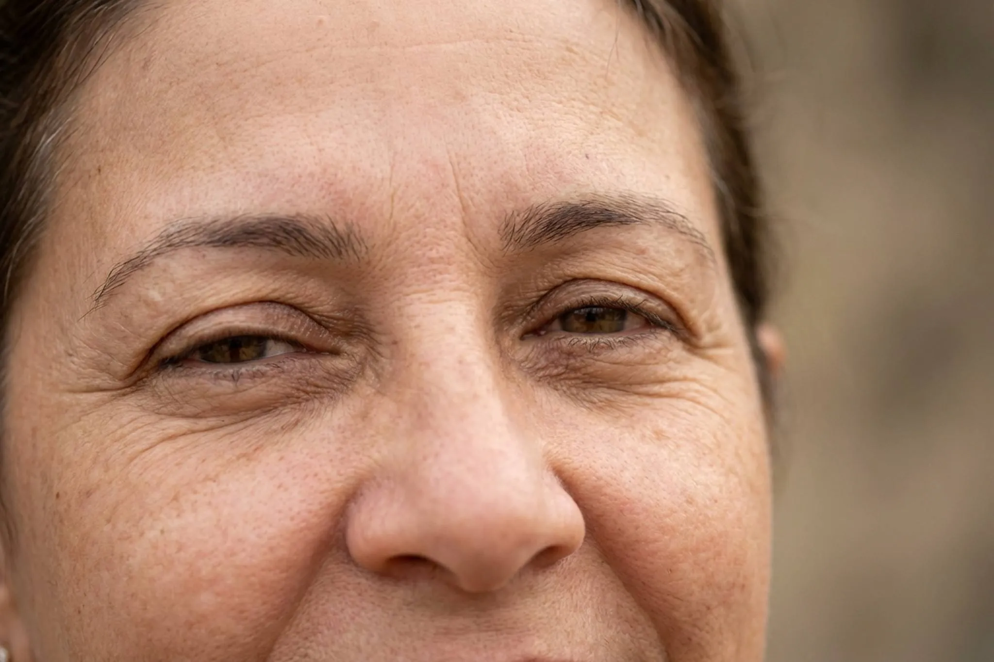
If you have noticed that one or both upper eyelids are progressively drooping, covering part of the pupil and making vision harder, you are likely facing eyelid ptosis. Commonly known as "droopy eyelid", this condition goes beyond an aesthetic concern — it is a functional problem that can significantly compromise your quality of life.
In my practice as a plastic surgeon in Londrina, Brazil, I have treated hundreds of patients with eyelid ptosis. Many arrived at my clinic thinking they only needed a blepharoplasty to remove excess skin, when the real problem was weakness of the levator muscle. This distinction is crucial: correct treatment depends on an accurate diagnosis. International patients who travel to Brazil for private surgery benefit from my extensive experience in both functional and aesthetic eyelid correction.
Eyelid ptosis occurs when the levator muscle — the muscle responsible for keeping the upper eyelid open — loses strength or detaches from its insertion into the tarsal plate. The result is an eyelid that descends below the normal position, potentially covering the pupil partially or completely. Unlike simple excess skin treated by blepharoplasty, ptosis involves muscular or aponeurotic dysfunction that requires specific surgical correction.
Many patients live with ptosis for years before seeking treatment. Some adapt by tilting the head back or constantly raising the eyebrows to compensate for the drooping eyelid. These compensations cause muscle fatigue and headaches, and mask the true severity of the problem. If this sounds familiar, there is a solution — and it is more accessible than you think.
The causes of eyelid ptosis: understanding why the eyelid has dropped
Eyelid ptosis can have several origins, and identifying the correct cause is essential to define the best surgical approach. In my experience, I classify the causes into four main groups:
Involutional ptosis (age-related)
By far the most common cause I encounter. Over the years the aponeurosis of the levator muscle — the tendinous structure connecting muscle to eyelid — stretches, thins, or partially detaches from the tarsal plate. The eyelid gradually droops over time. This type is usually bilateral, although one side is often more affected than the other.
Prolonged use of rigid contact lenses can accelerate this process, as repeated manipulation of the eyelid on insertion and removal contributes to aponeurotic stretching. Previous eye surgery, such as cataract surgery, can also trigger involutional ptosis.
Congenital ptosis
Present from birth, congenital ptosis occurs when the levator muscle does not develop properly during gestation. The muscle contains an abnormal amount of fibrous tissue rather than contractile muscle fibres, reducing its ability to elevate the eyelid. It can affect one or both eyes.
In children, congenital ptosis deserves special attention because it can cause amblyopia — "lazy eye" — if the drooping eyelid obstructs the visual axis during the critical period of visual development. In those cases, early surgical correction is essential. I do not operate on paediatric ptosis; I refer all children to Dr. Giovanni André Pires Viana (see below).
Neurogenic ptosis
Caused by problems in the nerves controlling the levator. The best known is paralysis of the third cranial nerve (oculomotor nerve), which in addition to ptosis can cause strabismus and pupillary dilation. Myasthenia gravis — an autoimmune disease affecting the neuromuscular junction — can initially manifest as eyelid ptosis, typically fluctuating (worse as the day progresses, better with rest). Neurogenic ptosis is not corrected in my practice and is referred to Dr. Giovanni Viana.
Mechanical and traumatic ptosis
Eyelid tumours, scars, chronic inflammatory processes and direct trauma to the eyelid or orbital region can cause ptosis through excessive weight on the eyelid or direct injury to the levator muscle and its aponeurosis. Previous eyelid surgery falls within this category as well.
During the consultation I perform a detailed examination to identify not only the presence of ptosis but its specific cause, as this determines the most appropriate surgical approach.
Ptosis versus blepharoplasty: the distinction many are unaware of
One of the most frequent confusions is between ptosis and dermatochalasis (excess skin on the upper eyelid). Although they can coexist — and often do — they are distinct conditions requiring different treatments.
What is dermatochalasis
Dermatochalasis is excess skin (and sometimes fat) on the upper eyelid. The extra skin hangs over the eyelid crease and may cover the eyelashes, but the eyelid margin itself remains in normal position. Treatment is upper blepharoplasty, which removes the excess skin and fat.
What is ptosis
In ptosis the problem lies at the eyelid margin: it descends below normal (which would be about one to two millimetres below the upper corneal limbus). Even if you remove all excess skin through blepharoplasty, the eyelid will remain droopy unless the levator muscle is repaired.
Why accurate diagnosis matters
I have seen patients who had undergone blepharoplasty elsewhere and were dissatisfied because "the eyelid was still droopy". The reason was simple: undiagnosed ptosis. Blepharoplasty removed the excess skin but did not correct the levator weakness.
It is very common in my practice to correct ptosis and perform blepharoplasty in the same surgical session. In patients over fifty, the combination of involutional ptosis and dermatochalasis is the rule, not the exception. I first correct the ptosis — plicating the levator — and then remove the excess skin. The result is a completely renewed appearance.
If you are unsure whether your problem is excess skin, ptosis, or both, an in-person consultation is the only reliable way forward. I take precise measurements of the palpebral fissure, levator function, and margin-reflex distance to determine what actually needs to be done.
Clinical assessment: how I diagnose and classify ptosis
Successful ptosis surgery begins with meticulous assessment. During the consultation I carry out a series of measurements and tests to determine not only severity but also the most appropriate technique.
Measurements I perform
- Margin-reflex distance (MRD1): the most important measurement. Distance between the upper eyelid margin and the light reflex at the centre of the pupil. Normal range: four to five millimetres. Mild ptosis: three to four; moderate: two to three; severe: below two.
- Palpebral fissure: the vertical distance between upper and lower eyelid margins. Normal: nine to twelve millimetres.
- Levator muscle function: I block the action of the frontalis with my thumb on the eyebrow and ask the patient to look down and then up. The excursion of the eyelid reflects muscle function. Good function (above 10–12 mm) is required to indicate levator plication. Fair: 8–12 mm. Poor: below 8 mm — referred to Dr. Giovanni Viana.
- Height of the eyelid crease: in involutional ptosis the crease is often higher than normal, suggesting aponeurotic disinsertion.
- Standardised photographic record: photos in multiple gaze positions (primary, up-gaze, down-gaze, lateral) are my main method of documentation and surgical planning. I do not use an intra-operative phenylephrine test.
What else I assess
I also examine the position of the eyebrows, since many patients with ptosis develop compensatory brow elevation that must be accounted for in surgical planning. I assess any associated dermatochalasis, facial symmetry, orbicularis function, corneal sensitivity and the tear film. I request ophthalmic review when appropriate, especially to exclude neurological causes.
Where myasthenia gravis is suspected, I request anti-acetylcholine receptor antibodies and, if necessary, electromyography. Excluding neurological causes before offering surgery is essential — in those patients the appropriate treatment is medical, not surgical.
The technique I perform: levator muscle plication only
In my practice I perform one single technique for eyelid ptosis correction: levator muscle plication of the upper eyelid. This indication is restricted to patients with good levator function — excursion greater than 10 mm on pre-operative assessment.
How levator plication works
Through an incision in the natural crease of the upper eyelid (the same used for blepharoplasty), I identify the levator muscle. I then perform a plication — folding the muscle on itself with internal PDS sutures — to shorten the effective length of the muscle and elevate the eyelid margin to the desired height. The internal PDS sutures are absorbed over six months; the skin sutures are removed between day 5 and day 7.
The incision is hidden in the eyelid crease and becomes practically invisible after healing. When indicated, I combine upper blepharoplasty in the same operation to remove excess skin.
Why I do not perform other techniques
Other techniques exist in the literature (conjunctivomullerectomy, frontalis sling with fascia lata, Müller's muscle resection, Fasanella-Servat). I do not perform any of those. Cases in which levator plication is not appropriate — primarily patients with poor levator function (< 10 mm), severe congenital ptoses and neurogenic ptoses — are referred to Dr. Giovanni André Pires Viana, formerly professor of periorbital plastic surgery at Escola Paulista de Medicina for many years and a Brazilian reference in complex ptosis. Honesty about what I do and what I do not do is part of ethical conduct.
Local anaesthetic with sedation: why the patient must be awake
Eyelid ptosis surgery is performed under local anaesthetic with sedation, not general anaesthesia. The reason is technical: during the procedure I need the patient to open and close the eyes, look up and look down, so that I can measure the eyelid height in real time and confirm the planned 2 mm over-correction.
Sedation provides comfort and relieves anxiety without abolishing the ability to follow commands intraoperatively. This intraoperative adjustment is the differentiator that allows reliable symmetry — something impossible under general anaesthesia, where the patient cannot move the eyes.
The 2 mm over-correction: not a mistake, it is planning
At the end of the operation, the operated eyelid is left 2 millimetres higher than normal — the so-called planned over-correction. This can look excessive in the immediate post-operative period and often prompts questions during the first few days. It is not a technical error: it is a calculation.
During the first month the muscle settles and the eyelid gradually descends, levelling with the contralateral eye at around 30 days. If I finished surgery with the eyelid already at the desired final height, over the month it would drop below target and the result would be under-corrected. The over-correction compensates for this predictable settling.
Understanding this is crucial so the patient accepts the initial "too high" look, which resolves on its own. Unnecessary post-operative anxiety is avoided by explaining it beforehand in the consultation.
The levator is diseased: there is no "definitive cure" in ptosis surgery
Patients should understand a key technical fact: the levator muscle in a ptotic eyelid is generally diseased — and that disease persists after surgery. Plication effectively shortens the muscle and corrects the droop at the time of surgery, but it does not treat the underlying cause (aponeurotic disinsertion, muscular dystrophy, tissue ageing).
As a consequence, a further operation may be needed later in life, typically between 5 and 15 years after the first correction, depending on the severity of the underlying muscle disease. For that reason we cannot speak of a "definitive cure" in eyelid ptosis surgery. What surgery offers is an effective and long-lasting correction — but not permanent in a lifetime sense.
This technical honesty is part of the consultation. Realistic expectations produce satisfied patients over the long term.
The operation step by step: how I perform ptosis correction
Here I describe the most common scenario in my practice: levator plication, frequently combined with upper blepharoplasty.
Anaesthetic and marking
I perform the surgery under local anaesthetic with sedation. Before starting, with the patient seated, I make precise markings on the eyelid: the eyelid crease, the amount of skin to be removed, and reference points for symmetry.
Incision and access
The incision is made in the natural crease of the upper eyelid, following the pre-operative marking. Where dermatochalasis is present, I remove the strip of skin and orbicularis. I then access the orbital septum and identify the levator aponeurosis and muscle.
Levator identification and plication
I identify the levator — in involutional ptosis often thinned, disinserted or elongated — and release it carefully. I carry out the plication of the muscle on itself using internal PDS sutures, shortening its effective length to bring the eyelid margin to the desired height. The critical point is placing the sutures in the exact position that will provide the appropriate elevation.
Intra-operative adjustment
Here lies the advantage of local anaesthetic: I ask the patient to open the eyes and compare the height and contour of both eyelids. I make fine adjustments to the sutures until the desired symmetry is achieved. This real-time adjustment is impossible under general anaesthesia and contributes substantially to superior results.
Closure
I close the incision with fine skin sutures that are removed between day 5 and day 7. The scar is hidden in the natural eyelid crease and becomes practically imperceptible within a few weeks.
The procedure lasts between 45 minutes and one and a half hours, depending on whether it is unilateral or bilateral and whether combined with blepharoplasty. The patient goes home the same day.
Post-operative recovery: what to expect
Recovery from eyelid ptosis surgery is generally smoother than patients imagine. Here is what you can expect at each stage:
First 48 hours
Expect swelling and bruising in the eyelid area — completely normal. I recommend cold compresses (ice wrapped in a clean cloth) for twenty minutes every hour during the first 48 hours. Keep the head elevated, including at night. The prescribed medication effectively controls discomfort, which is usually mild. Lubricating eye drops protect the cornea.
First week
Swelling peaks between day 2 and day 3 and then begins to subside. Bruises (purple patches) may extend to the cheek and gradually fade within ten to fourteen days. Skin sutures are removed between day 5 and day 7 in the clinic, quickly and with appropriate care.
Second to third week
Most of the oedema has subsided and the patient is socially presentable. The eyelid may show slight temporary asymmetry due to residual oedema — this is expected and resolves on its own. Light make-up can be used after sutures are removed.
One to three months
The result continues to refine. The scar in the eyelid crease matures and becomes increasingly discreet. Eyelid sensation, which may be altered in the first few days, normalises completely.
Final result
Between 3 and 6 months the final result establishes itself. The eyelid assumes its settled position, the contour looks natural and balanced, and patients report a significant improvement not only in appearance but also in the visual field. Time off work is typically 1 to 2 weeks. Oedema is slightly greater than with upper blepharoplasty alone (fibrosis can resorb more slowly).
Important after-care
- Avoid intense physical exertion for two to three weeks
- No contact lenses for at least two weeks
- Protect the eyes from sun with sunglasses
- Do not rub or scratch the eyes during recovery
- Use eye drops and ointment as prescribed
- Attend all follow-up appointments
Eyelid ptosis in children: I do not operate — I refer to Dr. Giovanni Viana
Congenital ptosis in children and babies requires a specific approach and sub-specialised expertise. I do not perform paediatric eyelid ptosis surgery in my practice. All paediatric patients who come to me for assessment are referred to Dr. Giovanni André Pires Viana, who served for many years as professor of periorbital plastic surgery at Escola Paulista de Medicina (São Paulo) and is a national reference for complex ptosis, including paediatric cases and those with poor levator function.
Why referral is the right course
Severe congenital ptosis can cause amblyopia ("lazy eye") if the eyelid covers the visual axis during the first years of life, when the brain is still developing vision. Such cases need very early surgery — often before age two — and techniques such as frontalis sling with autologous fascia lata or synthetic materials, which fall outside my usual adult levator plication practice.
Beyond the specific technique, surgery in children requires experience with paediatric general anaesthesia, intra-operative judgement without patient co-operation, and long-term follow-up (further procedures throughout childhood and adolescence are commonly needed). All of this justifies referral to a dedicated specialist.
What I do in the paediatric consultation
When parents bring a child with ptosis, I assess clinically, explain the degree of urgency (risk of amblyopia) and make a formal referral to Dr. Giovanni Viana with a report of my findings.
Risks and complications: transparency above all
As with any surgical procedure, eyelid ptosis correction carries risks I discuss openly with every patient. Transparency is the foundation of a trusting relationship.
Under-correction and over-correction
Eyelid height is measured in millimetres. A difference of just one millimetre is noticeable. Under-correction (eyelid lower than desired) and excessive over-correction (eyelid too high, making complete eye closure difficult) are the most common complications. Intra-operative adjustment under local anaesthetic greatly minimises this risk, but revision surgery may be required in a small percentage of cases.
Asymmetry
Achieving perfect symmetry between the two eyes is the greatest technical challenge of ptosis surgery. No human face is perfectly symmetrical, and small asymmetries are acceptable and natural. In more obvious cases a revision may be advised.
Lagophthalmos
Difficulty fully closing the eyes can occur in the first few days after surgery, especially during sleep. I prescribe ocular lubricants and night-time ophthalmic ointment. This usually improves as the oedema resolves and the tissues settle.
Dry eye
Patients with a pre-existing tendency to dry eye may experience temporary worsening after surgery. Pre-operative tear-film assessment is essential.
Haematoma and infection
Rare with proper technique and strict post-operative care. Stopping anticoagulants and anti-inflammatories before surgery reduces the bleeding risk.
In global medical literature, the revision rate in ptosis surgery is around ten to fifteen percent — higher than in most aesthetic procedures. I always inform patients of this because realistic expectations produce satisfying results. With intra-operative adjustment under local anaesthetic, my own revision rate is below that average.
Results: what ptosis correction can do for you
My patients often describe the results as transformative — and they are not exaggerating. The eyelid that used to cover part of the pupil returns to its natural position, revealing a gaze hidden for years.
Functional benefits
- Wider visual field: patients report seeing "more" — not because visual acuity has changed, but because the upper visual field is no longer obstructed.
- Elimination of compensatory fatigue: without needing to keep the eyebrows constantly raised to hold the eyes open, the frontal muscles relax and tension-type headaches disappear.
- Improved neck posture: patients who tilted the head back to compensate for ptosis return to a natural posture.
- More comfortable reading and screen use: activities that rely on a wide visual field become easier.
Aesthetic benefits
- A more open, youthful gaze: the eyelid at the correct height produces a rested, alert expression.
- Better facial symmetry: especially in unilateral ptosis, correction restores natural balance between the two sides of the face.
- Periorbital rejuvenation: when combined with blepharoplasty, the transformation of the gaze is complete.
- Renewed self-confidence: many patients report a significant improvement in self-esteem after surgery.
Durability
Eyelid ptosis correction is long-lasting. In involutional ptosis treated by levator plication, the result holds up for many years. In some patients there may be some recurrence over decades, which is natural as ageing continues. Even when this happens, a later revision is a simpler procedure than the original surgery — and may be needed 5 to 15 years later, given that the levator remains diseased.
My experience and approach in ptosis correction
I graduated from the State University of Londrina (UEL) and had the privilege of training with Professor Ivo Pitanguy. Over more than twenty years of practice I have performed more than eight thousand plastic surgeries, including hundreds of eyelid ptosis corrections. I am a full member of the Brazilian Society of Plastic Surgery (SBCP) and the American Society of Plastic Surgeons (ASPS).
Ptosis surgery requires a rare combination of deep anatomical knowledge, millimetric technical precision, and refined aesthetic judgement. The levator muscle and its aponeurosis are delicate structures that require careful handling. Success depends on very fine adjustments — literally one or two millimetres — that make all the difference in the final outcome.
My treatment philosophy
Every ptosis is different and there is no one-size-fits-all approach. In my practice I personalise the surgical plan for each patient based on the cause of the ptosis, levator function, age, expectations and associated conditions. That individualisation is what produces consistently good results.
I also believe in treating the periorbital region as a whole. Ptosis corrected alongside upper blepharoplasty, when indicated, gives a far more harmonious result than treating each problem in isolation. In the same way, if there is associated eyebrow descent, a brow lift can be combined for a complete result.
For patients seeking an integrated approach to facial rejuvenation, ptosis correction can be paired with facelift, fat grafting, facial fillers, or botulinum toxin, always respecting the anatomical particularities of each region.
Post-botox eyelid ptosis: not a surgical problem, reverses on its own or with eye drops
One of the most common internet searches is "eyelid ptosis after botox" — when botulinum toxin applied to the glabellar or frontal region diffuses into the levator muscle and causes a temporary droop. I do not perform surgery for post-botox ptosis, because it is not a surgical case.
Iatrogenic ptosis from botulinum toxin is a spontaneously reversible phenomenon. Improvement typically occurs between 4 and 12 weeks after onset, as the effect of the toxin wears off. During that waiting period an ophthalmologist can prescribe α-adrenergic eye drops (apraclonidine; phenylephrine at low concentration) that stimulate Müller's muscle and temporarily elevate the eyelid by 1–2 mm, easing the aesthetic discomfort while nature takes its course.
If you are looking for correction of real — structural, non-iatrogenic — ptosis, please refer to the previous sections of this page. If your ptosis started after a botulinum toxin injection, the correct course is to wait for spontaneous reversal, with or without eye drops on ophthalmic advice, and not to pursue surgery.
Do exercises work for eyelid ptosis?
The internet contains a lot of content about "exercises to strengthen the eyelid" as a solution for ptosis. In my practice, exercises do not correct eyelid ptosis when real levator muscle disease or aponeurotic disinsertion is present. The levator is a small, involuntary muscle and does not respond to strength training in the way larger skeletal muscles do.
Exercises may have a role in specific neurological contexts (facial palsy rehabilitation, for example), but that is a neurological or physiotherapy prescription, not a solution for ptosis caused by levator laxity. When patients arrive after months of trying exercises without result, the honest conversation is that surgery is the only intervention with proven efficacy.
Surgical contraindications: glaucoma, contact lens use and thyroid eye disease
Beyond the general contraindications of any surgery (coagulopathy, uncontrolled hypertension, active smoking), there are three relative contraindications specific to eyelid ptosis surgery that require careful assessment and that frequently lead me to decide not to operate:
- Glaucoma: raised intra-ocular pressure and chronic use of anti-glaucoma drops may affect the cornea and tear film, increasing the risk of post-operative complications (severe dry eye, corneal ulcer). I generally do not operate on patients with glaucoma.
- Active contact lens wearers: long-term use of rigid lenses is itself a known cause of involutional ptosis, and is also a risk factor for post-operative complications (lens intolerance after surgery, sensitised cornea). I generally avoid operating on patients who are actively wearing contact lenses.
- Thyroid eye disease (particularly Graves' orbitopathy): thyroid eye disease can cause eyelid retraction (the opposite of ptosis) and orbital changes that destabilise any eyelid surgical plan. I only consider surgery after stable endocrine control and a favourable ophthalmic review — or I refer.
Other situations in which I do not operate and refer: neurogenic ptosis (third nerve palsy, myasthenia gravis, post-stroke sequelae), ptosis with poor levator function, and all paediatric cases — all sent to Dr. Giovanni André Pires Viana.
Fees and insurance: private practice only
The fee for eyelid ptosis correction is agreed individually during the in-person consultation, after assessing levator function, degree of ptosis, laterality (unilateral or bilateral) and associated procedures (such as upper blepharoplasty). The initial consultation costs US$ 180. Postoperative follow-up appointments are included at no additional cost for one year after surgery. International consultation rates are listed in USD; payment is processed in Brazilian reais (BRL).
NHS and private medical insurance
In my practice, I operate only in private / self-pay arrangement. I do not work with the NHS nor with private medical insurance (BUPA, AXA Health, Vitality, Aviva, or others), even in cases with a documented functional component. If you need NHS or insurance cover for ptosis correction in the UK, the route is an IFR (Individual Funding Request) through your ICB where superior visual field obstruction is documented on Humphrey or Goldmann perimetry — and that is reviewed case by case by the NHS, not by my practice.
About the codes (ICD-10 and OPCS)
Eyelid ptosis is coded as ICD-10 H02.4. A corresponding OPCS procedure code exists for its surgical correction, frequently used in UK insurance claim requests. In my private Brazilian practice these codes are recorded in the clinical notes for documentation and for any post-payment reimbursement the patient may wish to claim independently from their insurer.
Unilateral versus bilateral ptosis: how I manage it
Most cases I see are unilateral (one eye). When the case is unilateral, I perform levator shortening on the affected side only — I do not manipulate the levator of the unaffected eye.
To improve visible symmetry between the two sides I may remove a small amount of skin from both upper eyelids (a small associated bilateral upper blepharoplasty), adjusting the eyelid crease height and overall appearance. This approach preserves the function of the healthy eye and only addresses any excess eyelid skin.
In true bilateral cases — both eyes with MRD1-documented ptosis — the plication is carried out on both sides, with individualised intra-operative adjustment for each eye. The 2 mm over-correction is applied bilaterally.
Frequently Asked Questions about Eyelid Ptosis
What is the difference between eyelid ptosis and excess eyelid skin?
They are different conditions. Eyelid ptosis is the drooping of the upper eyelid margin due to weakness of the levator muscle or disinsertion of its aponeurosis. Excess skin (dermatochalasis) is a fold of skin hanging over the eyelid crease, but the eyelid margin itself is in normal position. Treatments differ: ptosis requires muscle repair; excess skin requires blepharoplasty. The two conditions often coexist and can be corrected in the same procedure.
Is eyelid ptosis surgery performed under local or general anaesthesia?
Exclusively under local anaesthetic with sedation. That choice is technical, not a preference: during the procedure I need the patient to open, close and move the eyes up and down to confirm the planned 2 mm over-correction. Sedation keeps the patient comfortable without abolishing co-operation. I do not perform ptosis surgery on children (I refer to Dr. Giovanni A. P. Viana).
Which surgical technique do you use?
I perform levator muscle plication of the upper eyelid, indicated when levator function is good (excursion greater than 10 mm). Through an incision in the eyelid crease I fold the muscle on itself with internal PDS sutures to shorten it effectively. I do not perform conjunctivomullerectomy, frontalis sling, Fasanella-Servat or Müller's muscle resection — cases that require those techniques (poor levator function, severe congenital ptosis, neurogenic ptosis) are referred to Dr. Giovanni A. P. Viana.
Why did my eyelid look "too high" in the first few days?
That is expected and planned. At the end of surgery I leave the eyelid 2 millimetres higher than normal — the planned over-correction. Over the first month the muscle settles and the eyelid gradually descends to level with the contralateral eye at around 30 days. Without the over-correction the final result would be under-corrected.
How long does ptosis surgery take?
Unilateral correction takes 45 to 60 minutes. Bilateral, about 90 minutes. Combined with blepharoplasty the total time is 90 to 120 minutes. It is a day-case procedure — the patient goes home the same day.
Does ptosis correction leave a visible scar?
The incision is placed in the natural crease of the upper eyelid, hidden within the eyelid fold. Skin sutures are removed at 5–7 days; internal PDS sutures are absorbed over 6 months. After complete healing the line becomes practically invisible.
Can ptosis come back after surgery? Is there a definitive cure?
There is no "definitive cure". The reason is technical: the levator muscle is usually diseased (the underlying cause — aponeurotic disinsertion, dystrophy, ageing — is not removed by plication). A further operation may therefore be needed between 5 and 15 years later, depending on the severity of the muscle disease. The result is effective and long-lasting, but not permanent in a lifetime sense.
Does my private medical insurance cover ptosis surgery?
In my practice I operate only on a private / self-pay basis. I do not work with the NHS or with private medical insurers (BUPA, AXA Health, Vitality, Aviva and others), even in ptosis with a documented functional component. If you need NHS cover, the route is an IFR (Individual Funding Request) through your ICB with superior-visual-field obstruction documented on Humphrey or Goldmann perimetry — that is managed through the NHS, not my practice. ICD-10 (H02.4) and the equivalent OPCS code are recorded in the notes for any post-payment reimbursement the patient may wish to seek.
Do exercises work for eyelid ptosis?
Eyelid exercises do not correct structural eyelid ptosis when real levator disease or aponeurotic disinsertion is present. The levator is a small, involuntary muscle that does not respond to strength training like larger skeletal muscles. When patients have spent months on exercises without result, the honest conversation is that surgery is the only intervention with proven efficacy when it is correctly indicated.
I have glaucoma (or wear contact lenses, or have thyroid eye disease). Can I still have surgery?
Those three conditions are relative contraindications in my practice, and I generally do not operate on patients with glaucoma, active contact lens wearers, or patients with thyroid eye disease (particularly Graves' orbitopathy). The risk of post-operative complications (severe dry eye, eyelid instability, retraction) rises significantly. Each case is assessed individually in the consultation.
My ptosis is unilateral. Do you operate only on the affected side?
When eyelid ptosis is unilateral, I perform levator shortening only on the affected side. To improve visible symmetry I can remove a little skin from both upper eyelids (a small associated bilateral blepharoplasty), adjusting the eyelid crease. I do not manipulate the levator of the unaffected eye.
My child has ptosis. Do you operate on children?
I do not operate on children or babies with eyelid ptosis, because paediatric cases require sub-specialist expertise in periorbital plastic surgery. I refer all paediatric patients to Dr. Giovanni André Pires Viana, former professor of periorbital plastic surgery at Escola Paulista de Medicina and a Brazilian reference in complex ptosis. In the consultation I assess, explain urgency (amblyopia risk) and make a formal referral with a report.
Is there a non-surgical treatment for eyelid ptosis?
There is no structural, non-surgical treatment for ptosis. Temporary devices such as "eyelid crutches" attached to spectacles exist for cases where surgery is not possible. α-Adrenergic eye drops (oxymetazoline, apraclonidine) raise the eyelid by 1–2 mm temporarily — useful as palliation or for post-botox ptosis, but they do not replace surgery in structural ptosis. In ptosis due to myasthenia gravis, medical treatment of the underlying neurological disease may improve the condition.
Can I have eyelid ptosis correction and blepharoplasty at the same time?
Eyelid ptosis correction and upper blepharoplasty can be performed in the same surgical session when both conditions are present. Most adult patients with involutional ptosis also have excess upper eyelid skin. Both conditions are corrected through the same incision. The outcome is a more complete rejuvenation of the gaze.
How long until I see the final result?
The provisional result is visible within the first week, after suture removal. Residual oedema resolves within 4 to 6 weeks. The final result, with mature scar and stable eyelid contour levelled with the contralateral side, is established between 3 and 6 months. Typical time off work is 1 to 2 weeks.
Can botulinum toxin cause eyelid ptosis? Do you operate on post-botox ptosis?
Botulinum toxin can cause temporary eyelid ptosis when it migrates to the levator muscle after frontal or glabellar injections. I do not operate on post-botox ptosis — it is spontaneously reversible within 4 to 12 weeks, as the toxin wears off. During that period an ophthalmologist can prescribe α-adrenergic eye drops (apraclonidine, phenylephrine) that temporarily elevate the eyelid. Surgery is indicated only for structural, non-iatrogenic ptosis.
How should I prepare for ptosis surgery?
I request laboratory tests (full blood count, coagulation profile, glucose), a pre-operative cardiac review with ECG, and where relevant an ophthalmic assessment. Anti-inflammatories, aspirin, vitamin E and herbal remedies should be stopped 15 days before surgery. Smoking should be stopped for the same period. On the night before, an 8-hour fast is required.
Book via WhatsApp
If you have noticed that your eyelid is drooping, compromising your gaze or your vision, the next step is an in-person evaluation. During the consultation, I take all necessary measurements to diagnose the cause and degree of ptosis, and I explain exactly which technique will be most suitable for your case. My team is ready to assist you.
Learn more about the first consultation, the pricing, and the guidelines for pre-surgical preparation and post-operative recovery.
Recover your gaze! Book your consultation now.

Dr. Walter Zamarian Jr.
Plastic Surgeon in Brazil
Rua Engenheiro Omar Rupp, 186
Londrina - Brazil
ZIP 86015-360
Brazil
YouTube Channel: Dr. Walter Zamarian Jr.
Follow on Instagram: @drwalterzamarianjr
Follow on TikTok: @drwalterzamarianjr
Search on our website:
This page in other languages:
Portuguese (BR) | English (US) | English (UK) | Italian | French | Spanish